V.S. Anandarani
Associate Professor, Department of Anatomy, Sri Ramachandra Medical College, Chennai, India.
Corresponding Author: V.S. Anandarani, Associate Professor, Department of Anatomy, Sri Ramachandra Medical College, Chennai, India.
E mail: ranironeena@yahoo.co.in
Sir,
The luminal casts are useful as dry specimens in the museum and as teaching aids. During routine undergraduate teaching in the dissection hall when pancreatic ducts and their terminations are discussed, students become curious to see the herring-bone pattern of the pancreatic duct. Dissection of pancreatic ductal system is time-consuming and difficult because of the eye and throat irritation caused by formalin vapours. A luminal cast of ducts for easy and better demonstration is an appealing option.
In order to execute this option, two fresh pancreas specimens collected from the forensic department during autopsy were used for the procedure. The specimens were removed with duodenum and spleen. Bile duct was cut just above the first part of duodenum. Spleen was removed from the tail of the pancreas. After irrigating with water about 4 ml of latex solution was injected through the bile duct using a syringe and canula. The major and minor duodenal papillae were closed by suturing to prevent the leakage of latex solution. The specimens were put inside a solution of concentrated potassium hydroxide. The specimens were completely immersed in the solution. After 24 hrs when the soft tissues were corroded the casts were taken out and were cleaned thoroughly in tap water. After these procedures only one specimen of corrosion cast showing the branching pattern came out well. In the other specimen due to defective perfusion of the latex solution complete ductal system was not visible.
Corrosion cast revealed the typical ductal pattern of pancreatico-biliary tree. The smaller pancreatic ducts open into the main duct at regular angles resembling the “Herring bone pattern” (Figure 1).
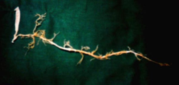
fig -1 Cast showing the ductal pattern of pancreas
The pancreas is a compound racemose gland analogus in its structure to the salivary glands. It is composed of two different types of glandular tissue which are however in intimate topographic association with each other. The exocrine tissue forms the main bulk of the pancreas. The pancreas is developed from dorsal and ventral pancreatic buds. The main pancreatic duct is normally derived from ventral and dorsal embryological buds of the pancreas. The accessory pancreatic duct is the major out flow tract of dorsal pancreatic bud in the embryo.
The corrosion cast showed that the main pancreatic duct after receiving smaller branches from tail, body, neck and postero-inferior parts of head joined the bile duct to form a common channel. The accessory pancreatic duct showed a communication with the main duct. It was placed in an anterior plane. It received branches from superior and inferior part of head of pancreas.
Casting techniques have been reported to be very useful to learn the complicated anatomy of ventricles, arterial supply of hilar bile duct, vascularisation of lips etc [1,2,3]. Dissection of pancreatic ductal system needs lot of time. In routine dissection, one has to be very close to the
specimen and so, formalin irritation to eyes and throat are unavoidable. Therefore, such cost-effective models are useful for demonstration as well as for retaining in museum.
References
-
Mittal PS, Khanwalker PG, Naik DC. Use of silicone gel to mould the cavities of hollow viscera. Journal of Anatomical Society of India 2009;58:201-2.
-
Gunji H, Cho A, Tohma T, Okazumi S, Makino H, Shuto K, et al. The blood supply of the hilar bile duct and its relationship to the communicating arcade located between the right and left hepatic arteries. Am J Surg 2006;192:276-80.
-
Crouzet C, Fournier H, Papon X, Hentati N, Cronier P, Mercier P. Anatomy of the arterial vascularisation of the lips. Surg Radiol Anat 1998;20:273-8.
Acknowledgement- The author expresses sincere thanks and gratitude towards Dr.Christilda Felicia, Former Director of Institute of Anatomy, Madras Medical College, Chennai. |