
| Case Report | ||
| Interesting neurological cause of scapular fracture | ||
| BK Bajaj, KS Anand
Department of Neurology, Postgraduate Institute of Medical Education & Research & Dr. RML Hospital, New Delhi-110001, India.
Case report A 42 year old male developed sudden onset drooping
of right shoulder while lifting himself up with his
right arm from squatting position. The patient Discussion Syringomyelia can lead to chronic neuropathic degenerative changes in joints usually shoulder and elbow. At presentation the X-ray appearance of Charcot's joint may be normal or show soft tissue calcification, intra-articular fractures, bone fragmentation and dislocation. Soft tissue swelling is an early finding. In the present case, only fracture of spinous process of scapula was evident and the patient did not have clinical and radiological evidence of Charcot’s arthropathy. It was the peculiar lack of pain and tenderness despite fracture of spine of scapula following relatively trivial trauma that led to further detailed assessment and discovery of syringomyelia. First descriptions of the destructive neuropathic arthropathy are by Mitchell and Charcot [1]. Mitchell and Charcot proposed that the arthropathy is result of damage to central nervous system trophic centres controlling nutrition of bones and joints. This theory is popularly known as the French theory. The theory proposed by Volkman and Virchow called the German theory, suggests that the arthropathy is the result of recurrent subclinical trauma that accumulates over years due to insensate joints. The reports of neuroarthropathy in patients who do not undergo repeated trauma or are bedridden prompted Brower and Allman to propose two theories, the neurotraumatic theory and the neurovascular theory [2]. The neurotraumatic theory suggests that repeated trauma resulting from loss of normal protective reflexes that prevent joints from exceeding the limits of normal range of motion, causes joint destruction. The neurovascular theory states that Charcot joint develops when sensory deficit disrupts the normal neurovascular reflexes around the joint resulting in hyperemia and activation of osteoclasts causing resorption of bone. Burcu Yanik and coauthors reported neuropathic arthropathy of shoulder and elbow joints in a patient with Arnold Chiari malformation with syringomyelia [3]. Neuropathic arthropathy may be the first manifestation of syringomyelia. Painless dislocation of shoulder joint without other changes are reported in literature [4]. As a result, an orthopaedician may be the first person of contact for a patient with syrinx. The present case differed from the reported cases in literature as there were no obvious features of Charcot arthropathy on clinical and radiological examination. However, MRI and technitium bone scan have been added to the armamentarium for investigation of Charcot’s arthropathy. These are more sensitive than a plain radiograph. The patient fractured his spine of scapula while doing the simple act of pulling himself up from sitting to standing position with the help of his arm. The sudden onset inability to abduct his arm with normal nerve conduction study and finding of cervical syrinx pointed towards recent development of fracture of spine of scapula with disruption of acromioclavicular joint as the cause of patient’s abrupt inability to abduct his right arm. The patient had lower motor neuron type of involvement in both the upper limbs (absent deep tendon reflexes in right upper limb with mild right hand grip weakness and diminished left upper limb deep tendon reflexes) and upper motor neuron type of involvement in lower limbs (bilateral brisk knee and ankle jerks). The patient did not have any difficulty to abduct his arm prior to the act of suddenly getting up from floor while taking support with his right arm. We believe that the inability to abduct the right arm was due to fracture of spine of scapula and not due to neurological cause. While patients with intramedullary lesion such as syringomyelia classically have early urinary and bowel dysfunction, it is not uncommon to see patients without significant involvement of bladder and bowel. This patient had bilateral brisk knee and ankle jerks with flexor plantar reflex. Typically, one expects plantar response to be extensor in such situations. It is well known clinical experience that plantars may be flexor in some of patients with pyramidal tract involvement. The impression of upper motor neuron type of involvement of lower limbs is made in the light of all the clinical symptoms and other signs elicited. This was further confirmed by MRI of the spine. Vinod kumar et al reported a case of fracture and displacement of body and neck of scapula in a post-traumatic paraplegic patient developing during the transfer from wheel chair to bed [5]. They reported that the patient was diagnosed to have cervicodorsal syrinx after it was realised postoperatively that the patient did not have the expected pain despite the fracture and subsequent operative intervention. They opined that the scapular fracture was a stress fracture consequent to underlying syringomyelia. To the best of our knowledge there is no other reported case in literature of scapular fracture in a patient with syrinx. In our case, there was no prior complaint to suggest syringomyelia. It was the detailed clinical examination which led to discovery of clinical signs of spinal cord involvement and confirmation of syrinx in the patient by MRI. The case underlines the need to be thorough in neurological assessment of patients presenting with upper body fractures particularly incident upon a trivial trauma and consider the possibility of syringomyelia as a causative factor.
References
|
||
 The patient was
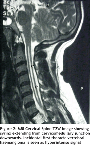 of
the upper limbs did not reveal any abnormality. The
patient’s cervico-dorsal Magnetic Resonance
Imaging (MRI) confirmed a syrinx extending from
cervico-medullary junction to the upper thoracic
segments (figure 2). Later, patient retrospectively
recalled that he had absent pain sensation for more
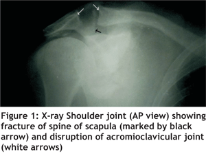
than 8 years in the right upper limb. X ray of the
shoulder joint did not reveal any definite changes
suggestive of Charcot’s arthropathy. Conservative management of fracture was done. Repeat
evaluation after around 8 weeks revealed persistent
fracture without any signs of healing.
of
the upper limbs did not reveal any abnormality. The
patient’s cervico-dorsal Magnetic Resonance
Imaging (MRI) confirmed a syrinx extending from
cervico-medullary junction to the upper thoracic
segments (figure 2). Later, patient retrospectively
recalled that he had absent pain sensation for more
than 8 years in the right upper limb. X ray of the
shoulder joint did not reveal any definite changes
suggestive of Charcot’s arthropathy. Conservative management of fracture was done. Repeat
evaluation after around 8 weeks revealed persistent
fracture without any signs of healing.