| Pictorial CME |
| Clinico-Sonographic Quiz |
Aruna Nigam, Sarita Choudhary, Chitra Raghunandan
Department of Obstetrics and Gynaecology, Lady Hardinge Medical College, New Delhi 110001, India.
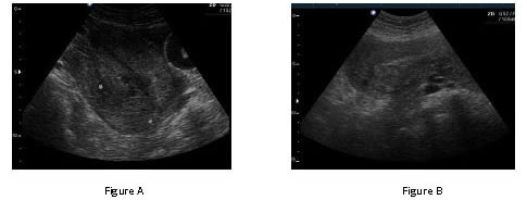
Corresponding Author: Aruna Nigam, Department of Obstetrics and Gynaecology, Lady Hardinge Medical College & associated hospitals, New Delhi-110001. India. A 24 year old primipara patient presented in the casualty with the complaint of retention of urine and heaviness in lower abdomen following delivery of male baby 6 days back at home. Per speculum examination shows rounded, oedematous mass in the vagina. Which of the following transabdominal ultrasonographic picture corroborates with her clinical findings?
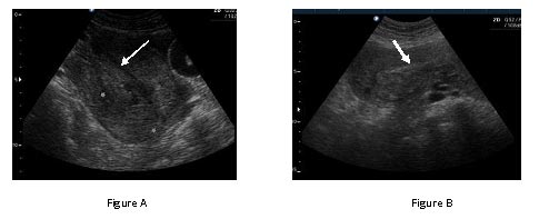
Answer to Clinico-Sonographic Quiz
Puerperal uterine inversion is the sequelae of mismanaged third stage of labour and usually presents with shock and haemodynamic instability in acute cases and with retention of urine and chronic pelvic pain in cases of subacute and chronic inversion. Active management of 3rd stage of labour is the main stay of prevention besides preventing anemia and promoting institutional deliveries. |